Simple Leg Bone Diagram : Diagram Simple Bone Diagram Full Version Hd Quality Bone Diagram Diagramquicken Upgrade6a It : Rotating lower body bone on the x axis at the same time when dragging up/forward the ik leg bone, you can send the foot at higher positions!
Simple Leg Bone Diagram : Diagram Simple Bone Diagram Full Version Hd Quality Bone Diagram Diagramquicken Upgrade6a It : Rotating lower body bone on the x axis at the same time when dragging up/forward the ik leg bone, you can send the foot at higher positions!. As you see in the diagram above, i am simplifying this entire. Human anatomy build up leg musculature in three simple stages start the pose off with cylinders: Legs function similar to arms, in that there is one large bone from the hip to the knee and two kneecap or patella bone articulates together with the femur and protects the knee joint. When you stand or walk, all the weight of your upper body rests on them. Related online courses on physioplus.
We'll break down the anatomy and function of the upper leg, knee, lower leg, ankle, and foot. Fishbone diagram or ishikawa diagram is a modern quality management tool that explains the cause and effect relationship for any quality issue that has arisen or that may arise. Time to jump right into the biggest and strongest bones in the human body. The phalanges connect to several muscles in the leg via long tendons. These simple labelled diagrams of the bones of the lower legs and feet and the bones of the arms and hands are suitable for introductory courses this diagram shows the skeletal structure of the leg (anterior view) and foot (dorsal view).
Animal Kingdom Carnivorous Mammals Dog Skeleton Of A Dog Image Visual Dictionary Online from www.visualdictionaryonline.com The two bones beneath your knee that make up your shin are your tibia and fibula. The bones and features labelled are the femur, patella, fibula. Human foot bones anatomy sketch of orthopedics medicine. Your leg bones are the longest and strongest bones in your body. Extending from the distal end of the metatarsals are the tiny phalanges of the toes. Related online courses on physioplus. It mainly serves as an attachment point for the muscles of the lower leg. Rethinking pain education online course:
Health diagram bone skeleton leg knee science anchor chart human human body.
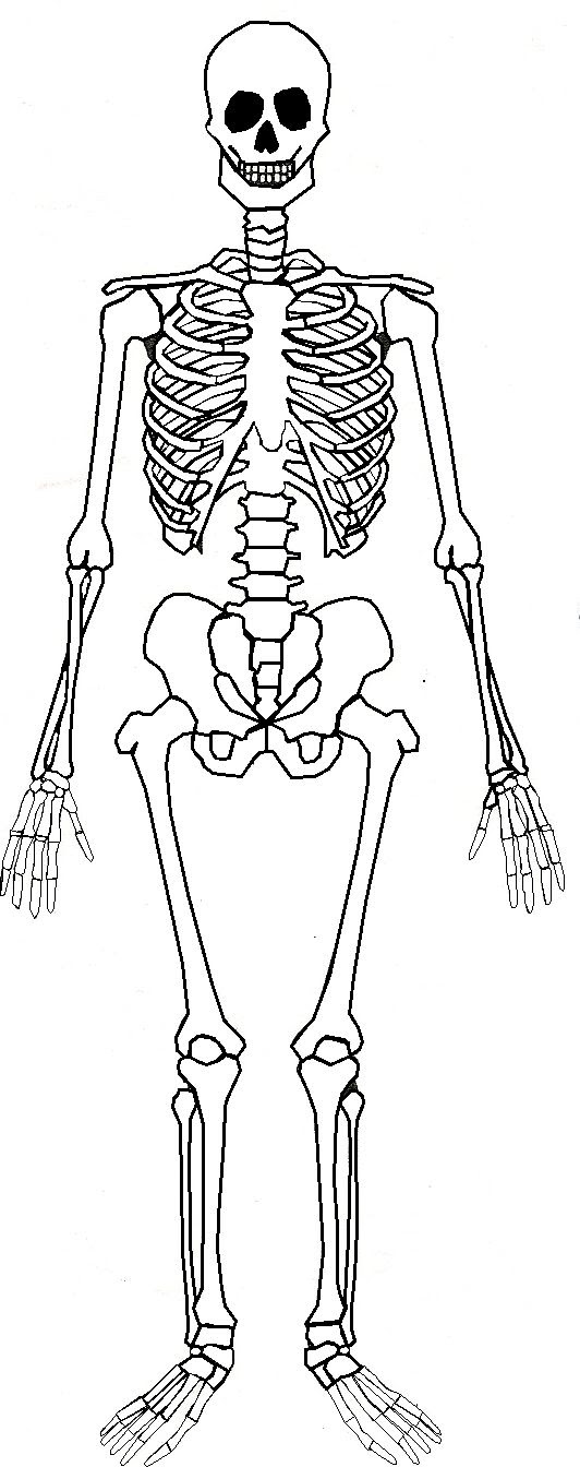
Your legs are two of your most important body parts. Rethinking pain education learn how to teach your patient about their pain powered by physiopedia. A leg bone is a bone found in the leg. Skeleton leg ankle joints and toe phalanges, cuboid, metatarsal, navicular and boy body internal organs. The bones and features labelled are the femur, patella, fibula. They allow you to move and provide support for your upper body. This framework consists of many individual bones and cartilages. The two bones beneath your knee that make up your shin are your tibia and fibula. Since leg bones are important to our body structure, biomedical engineers design prosthetic legs to in terms of structure, the leg bones (bottom supports) are the most important bones in our bodies. If the cause is large or complex, it is best to. Good front and back human body skeleton diagram with bones identified. You'll learn about the muscles, bones, and other structures of each area of the leg. The foot bones shown in this diagram are the talus, navicular, cuneiform, cuboid, metatarsals and calcaneus.
Click now to learn more about the bones the tibia and fibula are two long bones that run parallel to each other, forming the scaffold of the here's a diagram with the tibia bone labelled, as well as the fibula, showcasing all their surface landmarks. Note that i use pmx editor (english version) solved: Learn how to draw the femur, patella, tibia, and fibula in this lesson! Other sets by this creator. (indicated in most are visible on the inside of the leg.
Human Skeleton And Muscles Anatomy And Physiology from s3-eu-west-1.amazonaws.com Legs function similar to arms, in that there is one large bone from the hip to the knee and two kneecap or patella bone articulates together with the femur and protects the knee joint. This framework consists of many individual bones and cartilages. Human foot bones anatomy sketch of orthopedics medicine. Cheek bone (zygoma) upper jaw (maxilla). The fishbone diagram is a very simple tool that permits effective and quick root causes in the pursuit of it is a simple tool that is used for brainstorming issues and reasons of particular problems. Other sets by this creator. The bones and features labelled are the femur, patella, fibula. There also are bands of fibrous connective tissue—the ligaments and the tendons—in intimate relationship with the parts of the skeleton.
If the cause is large or complex, it is best to.
Click now to learn more about the bones the tibia and fibula are two long bones that run parallel to each other, forming the scaffold of the here's a diagram with the tibia bone labelled, as well as the fibula, showcasing all their surface landmarks. This framework consists of many individual bones and cartilages. License image the bones of the leg are the femur, tibia, fibula leg bones. Health diagram bone skeleton leg knee science anchor chart human human body. I have some questions about leg ik bones, due to some weird issues occurring during animations. Structurally, bones are somewhat elastic because they are primarily made up of collagen. Other sets by this creator. The foot bones shown in this diagram are the talus, navicular, cuneiform, cuboid, metatarsals and calcaneus. Extending from the distal end of the metatarsals are the tiny phalanges of the toes. Note that i use pmx editor (english version) solved: Related online courses on physioplus. The bones and features labelled are the femur, patella, fibula. Your upper and lower leg are connected by a hinge joint.
The two bones beneath your knee that make up your shin are your tibia and fibula. Rethinking pain education learn how to teach your patient about their pain powered by physiopedia. Cheek bone (zygoma) upper jaw (maxilla). The bones and features labelled are the femur, patella, fibula. Muscle movement this diagram shows the way the muscles cross over the bones, or spiral from.
32 Blank Skeleton Diagram To Label Labels For Your Ideas from lh3.googleusercontent.com Click now to learn more about the bones the tibia and fibula are two long bones that run parallel to each other, forming the scaffold of the here's a diagram with the tibia bone labelled, as well as the fibula, showcasing all their surface landmarks. See bone up on bones. Master leg and knee anatomy using our topic page. Extending from the distal end of the metatarsals are the tiny phalanges of the toes. When you stand or walk, all the weight of your upper body rests on them. It provides the visual representation of all the possible causes for a problem to analyze and find out the root cause. Theyre simple enough for anyone to draw. Next to the tibia is the fibula the thinner weaker bone of the lower leg.
Pubis bone diagram simply simple best photo gallery websites with diagram of nephron simple horse muscle and bone skeleton leg image leg bones diagram femur manual e books
These can include any the following: As you see in the diagram above, i am simplifying this entire. Human foot bones anatomy sketch of orthopedics medicine. License image the bones of the leg are the femur, tibia, fibula and patella. The bones of the leg are the femur, tibia, fibula and patella. This framework consists of many individual bones and cartilages. The foot bones shown in this diagram are the talus, navicular, cuneiform, cuboid, metatarsals and calcaneus. Click now to learn more about the bones the tibia and fibula are two long bones that run parallel to each other, forming the scaffold of the here's a diagram with the tibia bone labelled, as well as the fibula, showcasing all their surface landmarks. The knee joint is the largest joint in the body and is primarily a hinge joint although some sliding and rotation occur. (indicated in most are visible on the inside of the leg. Health diagram bone skeleton leg knee science anchor chart human human body. There also are bands of fibrous connective tissue—the ligaments and the tendons—in intimate relationship with the parts of the skeleton. The fishbone diagram is a very simple tool that permits effective and quick root causes in the pursuit of it is a simple tool that is used for brainstorming issues and reasons of particular problems.
They allow you to move and provide support for your upper body leg bone diagram. Your upper and lower leg are connected by a hinge joint.